
Stress granules (SGs) are cytoplasmic foci of RNA and proteins that form in cells under stress. Several research projects in the laboratory study properties and functions of these granules. SGs are best detected via microscopy using immunofluorescence (IF) staining for SG proteins or fluorescence in situ hybridization (FISH) of RNAs. In our laboratory we do lots of IF and FISH imaging of SGs and viral proteins. Here are some examples.
Sodium arsenite treatment induces stress granule formation (IF image):
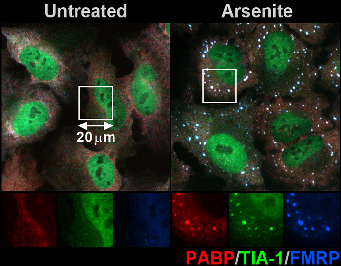
Above is a confocal microscopy image of U2OS cells untreated (left) or treated with sodium arsenite for 1 h (right) and stained with antibodies to PABP (red), TIA-1 (green), and FMRP (blue). Normally diffusely distributed in untreated cells, these proteins co-localize in arsenite-induced SGs (combination of all three colors gives bright white staining to granules).
Influenza A virus blocks stress granule formation in infected cells (FISH):

Above is a fluorescence microscopy image of A549 cells infected with influenza A virus and treated with sodium arsenite. Stress granules were stained with FISH probes for poly(A) tails of messenger RNAs that accumulate in SGs (red) and viral genome segments encoding M proteins (green). Nuclear DNA was stained with Hoechst dye.
Only uninfected cells that lack green staining form red SG foci, while infected cells do not form SGs (on the right panel green and blue channels were removed for clarity).
